Embryoscopy and Fetoscopy
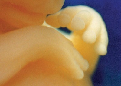
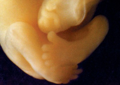
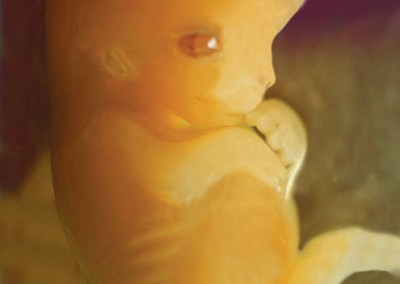
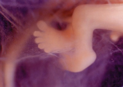
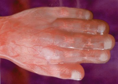
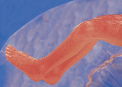
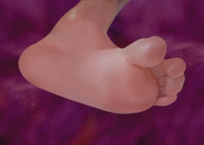
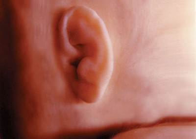
These cool color photos are obtained by using an embryoscope: a tiny camera the size of a pen tip. The e-scope, as I like to call it, is inserted through an incision in the abdomen and gently placed on the amniotic membrane. The results of this contact embryoscopy are spectacular!
Other Resources: If you would like to pick up some books and videos containing some truly amazing prenatal photography using the e-scope take a peak at these.
 From Conception to Birth : A Life Unfolds
by Alexander Tsiaras(Author)
Thanks go out to PFL and to Professor Andrzej Skawina (Collegium Medicum Jagiellonian University, Krakow) and Dr. Antoni Marsinek, MD (Czerwiakowski Gynecological and Obstetrics Hospital, Krakow) for some of the in utero images; UNC – Chapel Hill School of Medicine for their great resource and MP ULtrasound in Obstetrics and Gynaecology Europe Group.

